EGFR Osimertinib Resistance in NSCLC 2026: Mechanisms, Biomarkers and Combination Strategies
Linda
A 2026 translational review of osimertinib resistance in EGFR-mutant NSCLC, covering mechanisms, biomarkers, patient stratification, and next-line strategies.
A real translational-research case showing how molecular context can be converted into an actionable understanding of drug sensitivity, acquired resistance, biomarkers, patient stratification, and next-line development strategy.
This article is a topic-focused knowledge review of osimertinib resistance in EGFR-mutant NSCLC. It focuses on the biology, clinical evidence, resistance mechanisms, biomarkers, and treatment-development implications of this specific therapeutic problem rather than comparing AI research tools.
Method note: This article was built from a Noah AI Deep Research workflow that organized evidence from pivotal trials, regulatory sources, translational studies, and resistance datasets.
1. Molecular Context of EGFR-Mutant NSCLC
EGFR-mutant non-small-cell lung cancer is not one biologically uniform disease state. Drug response depends on the exact EGFR alteration, treatment line, co-alterations, tumor lineage, CNS involvement, and the evolutionary pressure created by previous EGFR tyrosine-kinase inhibitors.
The strongest clinical evidence for osimertinib centers on the common sensitizing variants EGFR exon 19 deletions and EGFR L858R. These variants define the core populations in first-line advanced disease, resected adjuvant treatment, and unresectable stage III disease after chemoradiotherapy. Acquired T790M belongs to a different clinical context: later-line disease after progression on an earlier-generation EGFR-TKI.
| Molecular / clinical context | Evidence base | Translational boundary |
|---|---|---|
| Untreated advanced NSCLC with exon 19 deletion or L858R | FLAURA randomized first-line evidence | Does not establish equal sensitivity for every uncommon EGFR alteration |
| Completely resected stage IB–IIIA NSCLC with exon 19 deletion or L858R | ADAURA adjuvant evidence | Applies to a selected resected population |
| Unresectable stage III disease after chemoradiotherapy | LAURA consolidation evidence | Does not replace definitive chemoradiotherapy |
| Acquired T790M after an earlier-generation EGFR-TKI | AURA3 later-line evidence | Represents a different treatment sequence from first-line osimertinib |
Uncommon EGFR variants, exon 20 insertions, and complex co-alteration patterns should therefore be interpreted at the allele level rather than grouped under a broad “EGFR-positive” label. Resistance data also change with treatment history: first-line osimertinib and later-line T790M-positive osimertinib create different selective pressures and different resistance landscapes.
2. EGFR Signaling and Osimertinib Mechanism
Activated EGFR signals through pathways including RAS–RAF–MEK–ERK and PI3K–AKT–mTOR, supporting proliferation, survival, and maintenance of the malignant phenotype. Sensitizing EGFR alterations can create a state of oncogene dependence, which explains why direct suppression of mutant EGFR can produce substantial tumor regression.
Osimertinib is a third-generation, irreversible, mutant-selective EGFR inhibitor. Sustained inhibition of mutant EGFR suppresses receptor autophosphorylation and downstream signaling. Its clinically demonstrated CNS activity is also important because intracranial control is a major component of disease management in EGFR-mutant NSCLC.
Resistance emerges when tumors restore signaling without relying on the same drug-sensitive EGFR configuration. That can occur through on-target EGFR evolution, bypass receptor activation, downstream pathway reactivation, or histologic and phenotypic transformation.
3. Molecular Contexts Associated With Drug Sensitivity
| Molecular context | Biological rationale | Evidence for sensitivity | Clinical relevance | Main limitation |
|---|---|---|---|---|
| EGFR exon 19 deletion | EGFR-dependent oncogenic signaling with favorable engagement by mutant-selective inhibition | Randomized evidence across FLAURA and related programs | One of the strongest established predictive contexts | Does not establish sensitivity of all exon 19 variants |
| EGFR L858R | Activating kinase-domain alteration producing EGFR dependence | Randomized evidence across metastatic and curative-intent settings | Core trial-validated osimertinib population | Response still varies with co-alterations and subsequent evolution |
| Acquired EGFR T790M | Preserves EGFR dependence while impairing earlier-generation inhibitors | AURA3 later-line randomized evidence | Established later-line predictive biomarker after earlier EGFR-TKI exposure | Not the expected dominant mechanism after first-line osimertinib |
| Selected uncommon EGFR variants | Variant-specific conformational and signaling effects | Functional and limited clinical evidence | Requires variant-level interpretation | Broad sensitivity cannot be inferred from EGFR mutation status alone |
4. Clinical Evidence of Osimertinib Response
Osimertinib has clinically established benefit across multiple EGFR-mutant NSCLC settings, but the magnitude and interpretation of benefit depend on treatment line, disease stage, comparator, and biomarker definition.
| Trial | Population / setting | Comparator | Major efficacy result | Key interpretation |
|---|---|---|---|---|
| FLAURA | Untreated advanced NSCLC with exon 19 deletion or L858R | Gefitinib or erlotinib | PFS 18.9 vs 10.2 months; OS 38.6 vs 31.8 months | Established first-line efficacy and important CNS activity |
| FLAURA2 | Untreated advanced EGFR-mutant NSCLC | Osimertinib monotherapy | Combination with platinum-pemetrexed improved PFS; HR 0.62 | Shows benefit from treatment intensification at the cost of greater chemotherapy-related toxicity |
| ADAURA | Completely resected stage IB–IIIA disease | Placebo | 5-year OS 88% vs 78% in the overall population | Supports prolonged adjuvant treatment in the studied common EGFR variants |
| LAURA | Unresectable stage III disease after chemoradiotherapy | Placebo | PFS 39.1 vs 5.6 months; HR 0.16 | Consolidation evidence after definitive chemoradiotherapy |
| AURA3 | T790M-positive disease after prior EGFR-TKI | Platinum-pemetrexed | PFS 10.1 vs 4.4 months; ORR 71% vs 31% | Validates T790M as a later-line osimertinib biomarker |
| COMPEL | Non-CNS progression on first-line osimertinib | Chemotherapy without continued osimertinib | PFS 8.4 vs 4.4 months; HR 0.43 | Supports continued EGFR suppression in a defined post-progression context |
5. Mechanisms of Acquired Resistance
Acquired resistance to osimertinib is not a single molecular event. It can arise through changes in the EGFR target itself, bypass signaling, downstream reactivation, lineage plasticity, or spatially distinct resistant clones. The apparent frequency of each mechanism depends on treatment line, assay, specimen type, and whether multiple progressing lesions are sampled.
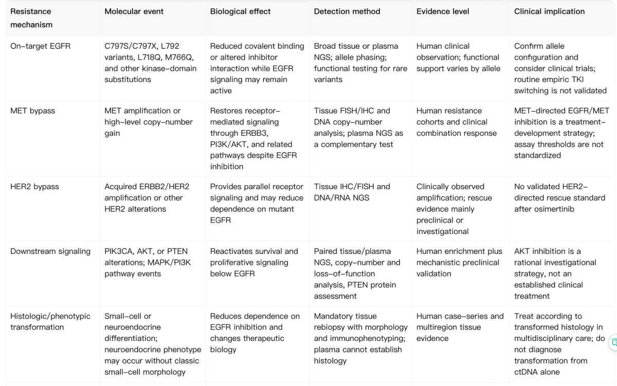
On-target EGFR resistance
On-target resistance preserves the EGFR-centered architecture but changes the receptor so osimertinib no longer suppresses it adequately. C797S is the best-known example because alteration of Cys797 can impair the covalent drug interaction. Less common kinase-domain substitutions can also affect binding, but rare variants require functional validation before they are treated as actionable.
MET and other bypass mechanisms
MET amplification is one of the most clinically supported bypass mechanisms. The resistant tumor may retain its founding EGFR alteration while becoming less dependent on EGFR as the sole signaling source. HER2 amplification and other receptor-level events can create similar parallel survival routes, although the strength of clinical evidence differs by mechanism.
Downstream reactivation
PI3K/AKT/PTEN and MAPK-pathway alterations can reactivate survival signaling below EGFR. These mechanisms are biologically plausible and supported by translational evidence, but detection does not automatically prove that a given pathway is the dominant resistance driver in a patient.
Histologic transformation
Small-cell or neuroendocrine transformation changes the therapeutic biology rather than simply adding one resistance mutation. Plasma ctDNA may detect accompanying genomic alterations, but it cannot establish histology. Tissue re-biopsy remains necessary when transformation is suspected.
6. Biomarkers for Sensitivity and Resistance
Biomarker testing should distinguish predictive biomarkers, resistance-enrichment markers, and exploratory findings. The best-established predictive contexts are baseline exon 19 deletion/L858R and, in a separate later-line setting, acquired T790M after progression on an earlier-generation EGFR-TKI.
| Biomarker / test | Sample type | What it indicates | When to use | Main limitation |
|---|---|---|---|---|
| EGFR exons 18–21 | Baseline tissue; plasma when tissue is unavailable | Defines EGFR-mutant disease and common sensitizing variants | At diagnosis and before stage-specific EGFR-directed treatment | Uncommon variants are not biologically equivalent |
| Plasma ctDNA | Plasma | Can detect sensitizing and acquired alterations | When rapid or minimally invasive testing is needed | Lower sensitivity for low-shedding disease, some fusions, copy-number events and transformation |
| Paired tissue re-biopsy | Progression tissue | Evaluates genomic resistance and histologic transformation | At progression when a systemic treatment change is being considered | Invasive and spatially limited |
| MET amplification testing | Preferably progression tissue, supplemented by plasma | Identifies a potential receptor-bypass state | At progression, especially when MET gain is suspected | Assays and thresholds are not standardized |
| EGFR C797S with allele phasing | Plasma or tissue | Supports on-target resistance interpretation | At progression when EGFR dependence may persist | Clinical meaning depends on configuration, clonality and coexisting mechanisms |
A negative plasma assay should be interpreted as a negative test result—not proof that no resistance mechanism exists. Tissue and liquid biopsy are complementary because plasma can miss spatially restricted amplifications, fusions, low-shedding disease, and histologic transformation.
7. Patient Stratification After Resistance
Post-osimertinib progression is best treated as a molecular and histologic reclassification problem. The relevant branches include persistent EGFR-dependent resistance, MET-driven bypass, other actionable signaling, mechanism-negative progression, and histologic transformation.
The key translational principle is to match the evidence burden to the proposed action. A candidate on-target mutation requires broad sequencing and often allele phasing; MET-driven disease requires robust amplification assessment; transformation requires tissue morphology; and mechanism-negative progression should trigger scrutiny of assay coverage, specimen quality, tumor shedding, and spatial heterogeneity.
8. Combination and Next-Line Development Strategies
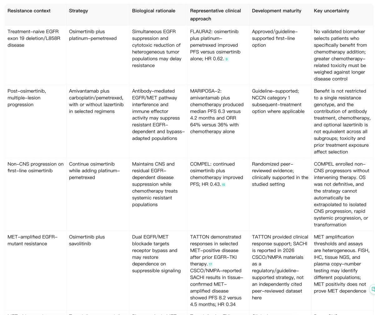
Combination development should follow the resistance state rather than assume that all progression requires the same intervention. The most mature strategies either intensify EGFR suppression, retain osimertinib while adding chemotherapy in a defined progression setting, or block a clinically supported bypass pathway.
Established and clinically supported approaches
FLAURA2 supports first-line osimertinib plus platinum-pemetrexed for selected patients who may benefit from greater treatment intensity. COMPEL supports continued osimertinib with platinum-pemetrexed in the studied non-CNS progression population. These are not interchangeable strategies and should not be extrapolated to every resistance pattern.
Biomarker-directed development
MET-directed combinations are the most developed example of mechanism-matched treatment after osimertinib resistance. Other approaches—including HER2-directed strategies, AKT inhibition, next-generation EGFR inhibitors, and kinase inhibitors for rare fusions—remain more dependent on trial-level evidence and should not be inferred from molecular detection alone.
9. What This Case Teaches Translational Researchers
A useful translational workflow does not stop at a list of molecular alterations. The stronger approach reconstructs an evolving tumor system and asks whether each observation is biologically and clinically linked to the treatment history.
Molecular alteration → pathway dependence → drug mechanism → response → resistance → biomarker → patient stratification → new therapeutic hypothesis.
Each link requires its own evidence: directionality, functional effect, cellular context, treatment context, and evidence strength. Human detection does not automatically establish causality. Functional rescue in a model does not automatically establish clinical benefit. A biomarker becomes useful only when the chain from observation to decision is sufficiently supported.
10. Bottom Line
Osimertinib remains clinically established across several common EGFR-mutant NSCLC settings, but post-treatment progression is molecularly heterogeneous.
MET amplification and on-target EGFR evolution are important resistance mechanisms, while downstream pathway activation, rare fusions, HER2 events, and histologic transformation require increasingly context-specific interpretation.
The key development opportunity is not simply to catalogue resistance mechanisms.
It is to connect:
resistance mechanism → biomarker → patient subgroup → evidence maturity → next-line strategy
and then determine which resistant dependencies can be suppressed reproducibly without excessive toxicity.
That is the step that turns molecular surveillance into translational strategy.
Build an Osimertinib Resistance Map with Noah AI
Build a source-linked resistance map across mechanisms, biomarkers, patient subgroups, and next-line strategies.
Use Noah AI to connect molecular alterations with pathway dependence, clinical evidence, resistance context, biomarker interpretation, and therapeutic-development options in one structured research workflow.
Free to use · Free credits included · No credit card required
Build a Resistance Map with Noah AI →